Pericarditis Constrictive Radiopaedia | Constrictive Pericarditis Radiology Reference Article Radiopaedia Org
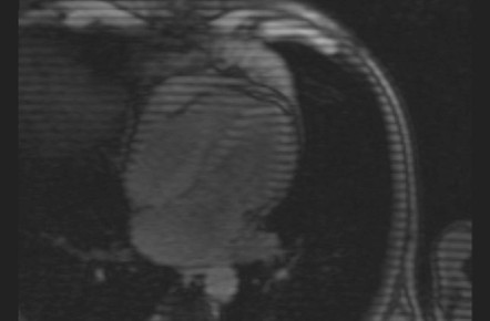
A Axial ECG-gated T1-weighted SE image shows pericardial thickening arrows which is most visible near the right ventricle. CT features are suggestive of constrictive pericarditis.
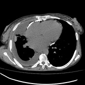
This is a case of pericarditis diagnosed on computed tomography.
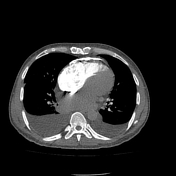
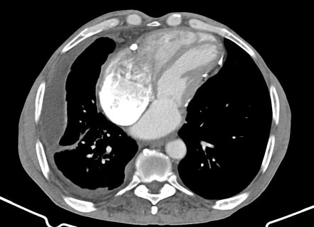
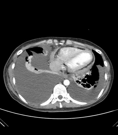
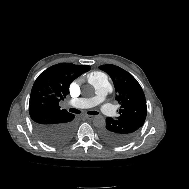
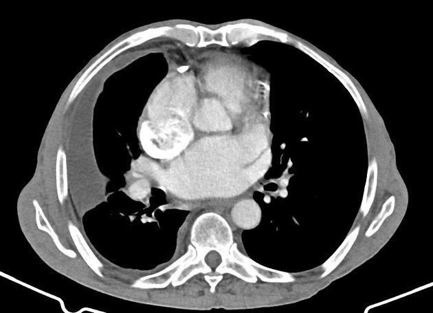
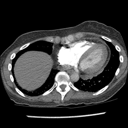
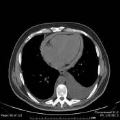
Pericarditis constrictive radiopaedia. There is heterogeneously enhancing diffuse nodular pericardial thickening consistent with an infectiveinflammatory pericarditis. Become a Gold Supporter and see no ads. Features of cardiac failure are present like flattening of the interventricular septum dilatation of inferior vena cava and presence of bilateral mild pleural effusion.
In the US and most other developed countries the most common causes are prior surgical violation of the pericardium mediastinal. A dilated right atrium dilated hepatic veins dilated. CT chest showed calcified thickened pericardium with tubular morphology of both ventricles and biatrial dilatation - consistent with calci.
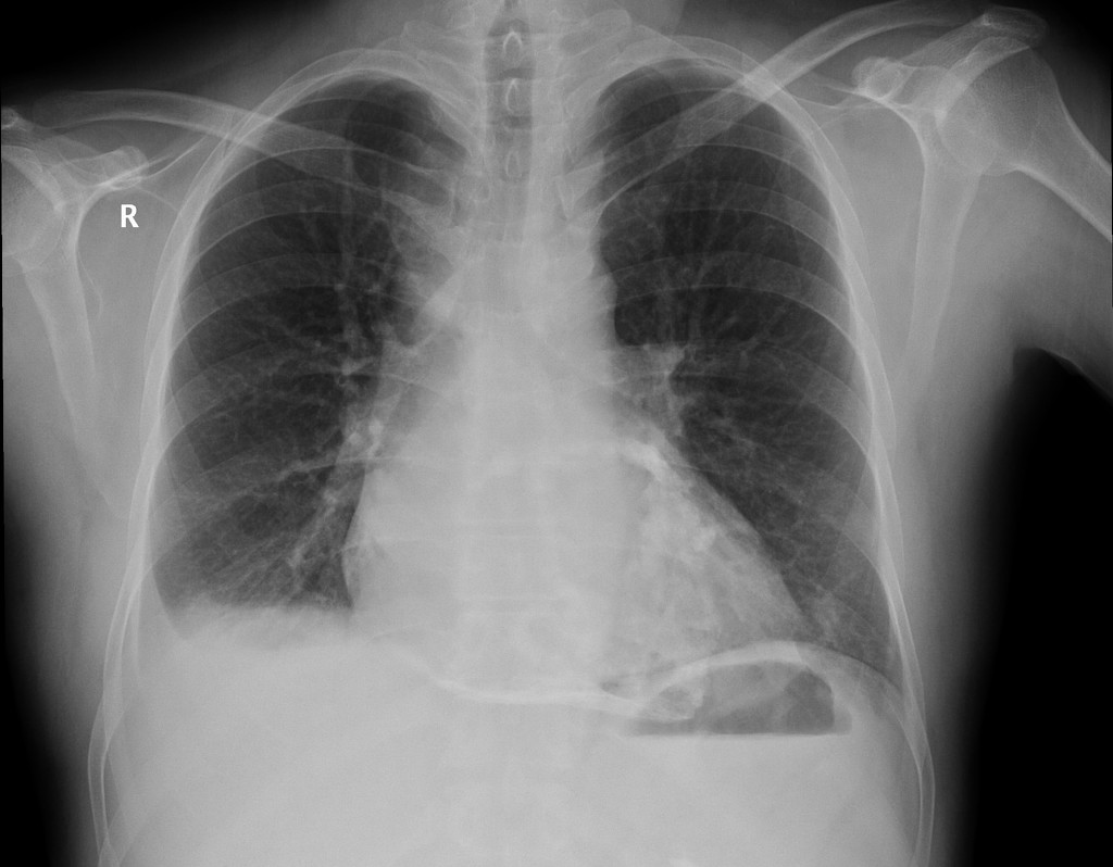
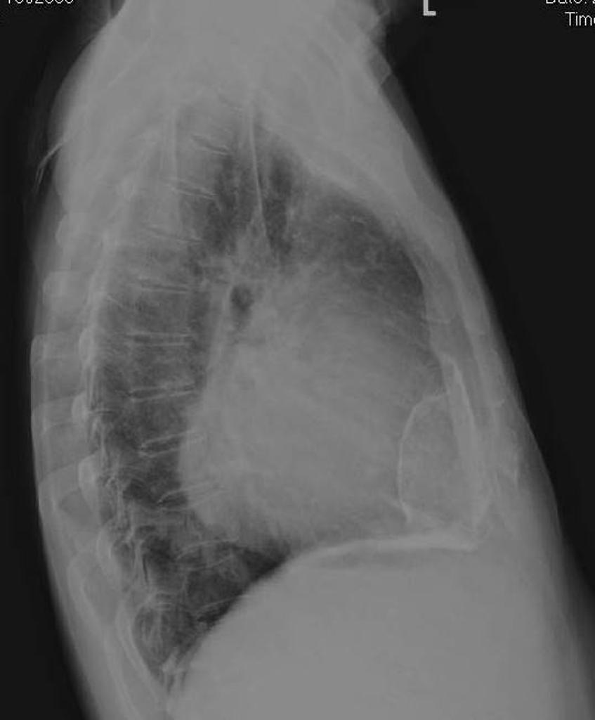
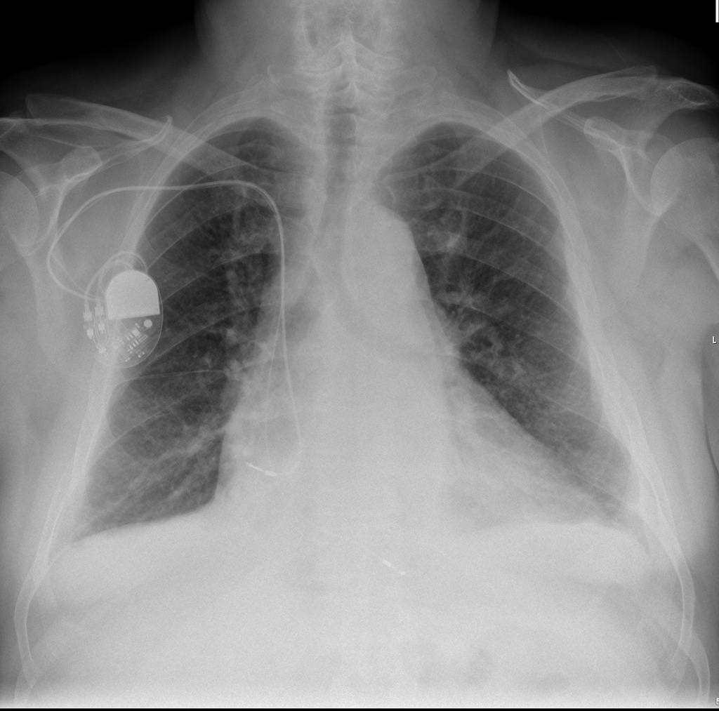
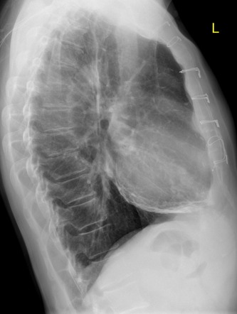
Distortion of the normal cardiac contour indicates constrictive physiology due to thickening and calcifications of the pericardium. It often has a characteristic distribution involving th. The heart is not.
The presence of right pleural thickening with effusion and par. Imaging signs that suggest heart failure especially right-sided dysfunction include. Infection remains the most common cause of pericarditis accounting for two-thirds of cases.
The presence of pericardial calcifications in conjunctions with signs of right sided heart failure is consistent with constrictive pericarditis. Axial C arterial phase. The right ventricle has a narrow tubular shape secondary to pericardial constriction.
The constellation of CT scan x-ray and echo-cardiogram findings in addition to. Constrictive pericarditis in a 65-year-old man who had had symptoms of right heart failure for 3 months. Noninfectious causes account for one-third of cases.
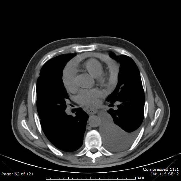
Noninfectious causes account for. In this case the patient was not complaining of chest pain the presentation was for shortness of breath which is the result of bilateral pleural effusions as a complication of pulmonary h. Calcification occurs among half of the patients with constrictive pericarditis.
Calcific thickening of the pericardium is seen which in a proper clinical setting is suggestive of constrictive pericarditis. This disorder must be considered in the differential diagnosis for unexplained heart failure particularly when the left ventricular ejection fraction is preserved. The thickest pericardial rind measures 13 cm.
This is a case of pericarditis diagnosed on computed tomography. In constrictive pericarditis CT classically shows thickening 4 mm of the pericardium most commonly anteriorly and pericardial calcifications. This causes interstitial edema in the right lung.
Finish Not needed End of previous page. 70 year old male with past history of pulmonary Kochs came with increasing breathlessness and chest pain. Infection remains the most common cause of pericarditis accounting for two-thirds of cases.
Pericardial calcification in the setting of heart failure is highly suggestive of constrictive pericarditis. Radiopaedia is free thanks to our supporters and advertisers. Constrictive pericarditis CP is a form of diastolic heart failure that arises because an inelastic pericardium inhibits cardiac filling.
Become a Gold. B Axial T1-weighted SE image obtained at a level slightly caudad to that in. When the pericardium becomes thickened or fibrosed it becomes less compliant and prevents adequate diastolic filling of the ventricles a condition known as constrictive pericarditis CP.
The patients echocardiogram showed an ejection fraction of approximately 50 left ventricular impairment bi-atrial enlargement and grade I mitral regurgitation. However radiographical evidence of constrictive pericarditis should also be accompanied by evidence of heart failure. Risk factors for the development of CP include prior cardiac surgery and radiation therapy.
Radiopaedia is free thanks to our supporters and advertisers.
Constrictive Pericarditis Radiology Case Radiopaedia Org

Constrictive Pericarditis Radiology Case Radiopaedia Org

Constrictive Pericarditis Radiology Case Radiopaedia Org

Constrictive Pericarditis Radiology Reference Article Radiopaedia Org

Constrictive Pericarditis Radiology Reference Article Radiopaedia Org
Constrictive Pericarditis Radiology Case Radiopaedia Org
Constrictive Pericarditis Radiology Case Radiopaedia Org

Calcific Constrictive Pericarditis Radiology Case Radiopaedia Org

Calcific Constrictive Pericarditis Radiology Case Radiopaedia Org
Constrictive Pericarditis Radiology Reference Article Radiopaedia Org
Constrictive Pericarditis Radiology Case Radiopaedia Org

Constrictive Pericarditis Radiology Reference Article Radiopaedia Org
Constrictive Pericarditis Radiology Case Radiopaedia Org
Pericarditis Radiology Reference Article Radiopaedia Org
Constrictive Pericarditis Radiology Case Radiopaedia Org
Constrictive Pericarditis And Pericardial Effusion Radiology Case Radiopaedia Org
Constrictive Pericarditis Radiology Reference Article Radiopaedia Org
Constrictive Pericarditis Radiology Reference Article Radiopaedia Org
